Module N7 · Topic 1 of 4
Indications, anatomy, and setup
The pulmonary artery catheter is a balloon-tipped venous catheter that floats into the pulmonary artery and reports pressures from the right heart, a stand-in for left-heart filling, and cardiac output. Using it well starts by knowing when it is indicated, what its numbers really mean, and how the setup makes those numbers trustworthy.
When it is indicated
There are no absolute, must-place indications. The catheter earns its place when the hemodynamic question is genuinely unclear: undifferentiated or multifactorial shock, monitoring cardiac function during and after cardiothoracic surgery, guiding treatment in advanced cardiogenic shock, sorting out and optimizing right heart failure and pulmonary hypertension (where right heart catheterization is the gold standard for diagnosis), sampling mixed venous oxygen saturation, and titrating mechanical circulatory support.
It is also important to recognize the limits of a PA catheter. The catheter gives static filling pressures, which do not in and of themselves predict fluid responsiveness. There is no single cardiac output or resistance number to chase, since an index that is fine for one patient leaves another in shock. Randomized trials show no mortality benefit, but more recent observational data suggests that use of PA catheters to guide care in certain populations is beneficial. The value is diagnostic clarity in the right patient, not a target to treat.
When not to place one
Some anatomy makes placement more difficult, changing the relative risk of harm caused by catheter placement. A tricuspid or pulmonic prosthesis, a valvular vegetation, or a right-sided mass or clot should lead to reassessment of the true risk:benefit of catheter placement. An existing left bundle branch block (LBBB) deserves special caution since catheter placement often provokes a transient right bundle branch block as it crosses the right ventricle. In a patient who already has a LBBB, this can result in unintended complete heart block. While an existing LBBB does not preclude PA catheter placement, it does suggest that equipment for emergent pacing should be readily available during placement.
Most other problems (arrhythmia, right bundle branch block in about 6 percent, line infection, pneumothorax on insertion, thrombus) come from the line itself, so plan to remove the catheter as soon as possible. Leaving a line in without acceptable reason is dangerous and frequently leads to unintended, harmful consequences.
The complication to respect
The complication to respect most is pulmonary artery rupture. While exceedingly rare, the complication carries a very high risk of fatality should it occur. The habits that prevent it are simple and non-negotiable:
- Watch the waveform during balloon inflation and stop the moment the wedge tracing appears.
- Never inject more than 1.5 mL of air into the balloon.
- Never inject fluid through the balloon port.
- Never leave the balloon inflated for a prolonged period of time (more than about fifteen seconds), and let the balloon deflate passively.
Know the catheter by its ports
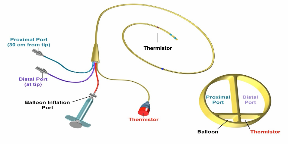
The catheter is easiest to learn by its lumens. Most are quadruple-lumen, and the color tells you the job:
| Port | Color | Ends at | Use |
|---|---|---|---|
| Proximal infusion | White or clear | 30 cm, right atrium | fluids and drugs |
| RA / injectate | Blue | 30 cm, right atrium | RA (CVP) pressure, injectate for cardiac output |
| Distal / PA | Yellow | tip, pulmonary artery | PA pressure and mixed venous sampling |
| Thermistor | Red | ~4 cm from distal tip | senses temperature for thermodilution |
A balloon surrounds the tip. The syringe for inserting air into the balloon usually withdraws to a maximum 1.5 mL, which is the built-in safety limit. Also note the dashes along the outside of the catheter itself. Thin markings on the catheter represent 10 cm increments of depth. Thicker markings represent a 50 cm depth increment.
Zero and level before you trust a number
Before any pressure means anything, the transducer has to be zeroed to atmospheric pressure and leveled to the phlebostatic axis. Level it even a few centimeters off and every pressure afterward is wrong. Run a trifurcated, pressurized saline bag at 300 mmHg, transduce the RA (blue) and PA (yellow) lines continuously so you catch an unintentional wedge or a drop back into the ventricle. For reference, the normal values you are checking against:
| Site | Normal pressure |
|---|---|
| RA / CVP | 2 to 6 mmHg |
| Right ventricle | 15 to 25 / 2 to 6 mmHg |
| Pulmonary artery | 15 to 25 / 8 to 15 mmHg (mean 10 to 20 mmHg) |
| Wedge (PAOP) | 6 to 12 mmHg |
| Cardiac index | 2.5 to 4 L/min/m2 |
Zeroing: what the number is measured against
Zeroing means setting the reference pressure from which every later pressure is measured. That reference is atmospheric pressure, and the choice is not arbitrary. The absolute pressure inside the right atrium is roughly one atmosphere plus a few mmHg, and nothing clinically useful lives in that number. What distends a chamber and what drives flow is a pressure difference. Referencing the transducer to atmosphere turns the displayed value into that difference, which is why a CVP of 5 mmHg means the same thing in Denver as it does at sea level even though the ambient barometric pressure differs by more than 100 mmHg.
Mechanically, the transducer is a strain gauge. It converts deflection of a thin diaphragm into a voltage, and the monitor converts that voltage into a pressure. Zeroing tells the monitor which voltage corresponds to no pressure difference across the diaphragm. Without it, any electrical offset in the gauge, the cable, or the amplifier is added to every subsequent reading as a fixed constant. That is the dangerous failure mode: the waveform still looks entirely normal, the morphology is preserved, and only the absolute numbers are wrong. A miscalibrated zero does not announce itself.
One caveat follows directly from the choice of reference. A pressure referenced to atmosphere is not a transmural pressure, because the chamber sits inside a thorax whose pressure is not atmospheric. That is why filling pressures are read at end expiration, the point in the respiratory cycle when pleural pressure comes closest to atmospheric and the referenced number comes closest to the distending pressure the ventricle actually sees.
To zero, position the transducer at the intended reference level first, then turn the stopcock nearest the transducer off to the patient and open to air, replacing the non-vented cap with a vented one so the diaphragm sees room air. Confirm the tracing falls to a flat line, select zero on the monitor, and confirm the displayed value reads 0 mmHg. Close the stopcock to air, replace the cap, and confirm a normal pressure waveform returns before you believe anything on the screen. Re-level after any change in patient position. Re-zero after any cable, transducer, or monitor change, at least once a shift, and any time a number is not believable.
Leveling: where zero sits in the body
Zeroing establishes what zero pressure means electrically. Leveling establishes where in the patient that zero sits. The two are separate steps and a perfect zero at the wrong height is still a wrong number.
The reason is hydrostatic. The catheter, the tubing, and the transducer form one continuous fluid column, so the transducer reports the pressure at its own height rather than at the height of the chamber being measured. Each centimeter the transducer sits below the reference point adds about 0.74 mmHg to every reading, and each centimeter above subtracts the same. A transducer taped 10 cm too low overreads by roughly 7 mmHg, which is the entire normal range of right atrial pressure. This is why sloppy leveling can render every subsequent measurement not merely imprecise but clinically misleading.
The phlebostatic axis, conventionally the fourth intercostal space at the mid-axillary line, is exactly that: a convention. It is an external landmark chosen to approximate the level of the right atrium, and more specifically to approximate the point of hydrostatic indifference, the level in the circulation where pressure changes least as body position changes. Adopting a single agreed reference point is what makes one patient's RA pressure comparable to another's, and what makes today's wedge comparable to yesterday's.
The convention is not settled and it is worth knowing why. The mid-axillary landmark is anatomically imprecise, particularly in obesity and in chest wall deformity, and the true point of hydrostatic indifference shifts with posture and with volume state, since the leg veins hold enough blood that upright and supine positions do not share one indifference point. Partly for this reason the mid-thoracic (Winsor/Burch) level, defined at the fourth intercostal space in the transverse plane and at mid-chest in the anteroposterior plane, has been adopted in the pulmonary hypertension literature. The practical rule is unchanged: pick the reference your institution uses, measure supine, re-level whenever the bed height or the patient's position changes, and keep it identical between serial measurements.
References
- Sandham JD, Hull RD, Brant RF, et al. A randomized, controlled trial of the use of pulmonary-artery catheters in high-risk surgical patients. N Engl J Med. 2003;348(1):5-14. doi:10.1056/NEJMoa021108.
- Wheeler AP, Bernard GR, Thompson BT, et al; National Heart, Lung, and Blood Institute Acute Respiratory Distress Syndrome (ARDS) Clinical Trials Network. Pulmonary-artery versus central venous catheter to guide treatment of acute lung injury. N Engl J Med. 2006;354(21):2213-2224. doi:10.1056/NEJMoa061895.
- Harvey S, Harrison DA, Singer M, et al. Assessment of the clinical effectiveness of pulmonary artery catheters in management of patients in intensive care (PAC-Man): a randomised controlled trial. Lancet. 2005;366(9484):472-477. doi:10.1016/S0140-6736(05)67061-4.
- Binanay C, Califf RM, Hasselblad V, et al. Evaluation study of congestive heart failure and pulmonary artery catheterization effectiveness: the ESCAPE trial. JAMA. 2005;294(13):1625-1633. doi:10.1001/jama.294.13.1625.
- Garan AR, Kanwar M, Thayer KL, et al. Complete hemodynamic profiling with pulmonary artery catheters in cardiogenic shock is associated with lower in-hospital mortality. JACC Heart Fail. 2020;8(11):903-913. doi:10.1016/j.jchf.2020.08.012.
- Kanwar MK, Blumer V, Zhang Y, et al. Pulmonary artery catheter use and risk of in-hospital death in heart failure cardiogenic shock. J Card Fail. 2023;29(9):1234-1244. doi:10.1016/j.cardfail.2023.05.001.
- Bertaina M, Galluzzo A, Rossello X, et al. Prognostic implications of pulmonary artery catheter monitoring in patients with cardiogenic shock: a systematic review and meta-analysis of observational studies. J Crit Care. 2022;69:154024. doi:10.1016/j.jcrc.2022.154024.
- Ortega-Hernández JA, Chacon-Lozsan FJ, Baldetti L, et al. Pulmonary artery catheter monitoring in cardiogenic shock: a systematic review and meta-analysis. Shock. 2026;65(4):648-659. doi:10.1097/SHK.0000000000002784.